Microtoming is an experimental technique which slices an sections of a sample for microscopic examination, using a light or electron microscope. Experimentally, the preparation technique varies depending on the sample, with different knives used to prepare different thickness's of section. The layers of the PoreXpert unit cell can be visualised in the 2D cell inspector in the xy and xz - plane. The thickness of an experimental section from a microtome can be as small as 100 nm, however the technique has limitations if you want to reconstruct the three dimensional structure as, the microtoming always loses the thickness of the knife, and this can be much thicker than the sections obtained.
The microtoming algorithm uses the unit cell output from the simplex and performs a microtoming operation in the xy, xz and yz plane. Each layer in the microtoming algorithm is sliced into 10 sections, so when dealing with cuboidal unit cells, there will be differences between the xy, xz and yz plane on the histogram. The next screen shows the output from the microtoming screen for a cuboidal sample.

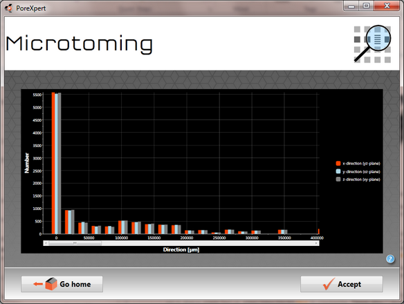
Microtoming display for a cuboidal 15 * 15 *10 unit cell. The graph shows small changes in the x - y and z directions shown in the three colours.
The microtoming graph can be wider than the screen so you may need to scroll to the right to view all of the detail from the microtoming operation.
As the Auto Cluster Ratio calculation is new for PoreXpert version 2.0, it has not yet been propagated into other simulations such as microtoming, so you will continue to see an artificial spike at the top of the size distribution. The identification of clusters allows this spike to be spread down through the other sizes - see our void size and permeability validation for a specific example of that.